ARISE Veterinary Center - Queen Creek

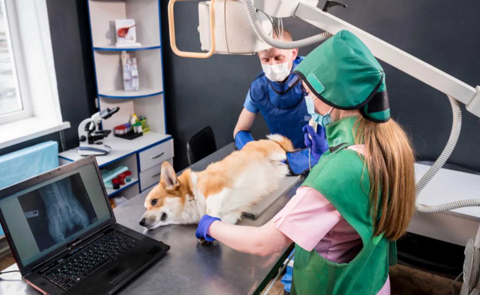
Diagnostic imaging involves the use of radiography and fluoroscopy (x-rays), ultrasonography, computed tomography (CT), and magnetic resonance imaging (MRI) to obtain information about the anatomy of inner organs of the chest and abdomen, as well as complex regions such as the head (brain), neck, and spine with minimal risk to the patient. These modalities complement each other and are considered non-invasive diagnostic techniques that provide information that cannot be obtained on physical examination or bloodwork. In addition, further information about the abnormalities identified can be obtained by using imaging modality techniques to guide sampling procedures such as fine needle aspirates and/or biopsies. All the information obtained can help the clinician reach a diagnosis and aids in determining the next step required to manage a patient’s illness
Tests and Procedures We Offer
We offer a number of advanced diagnostic and therapeutic options, including but not limited to:
Endoscopy: Endoscopy is a procedure in which a camera is inserted into an area of the body and images are taken. Endoscopy can be performed in the stomach, small and large intestine, bladder, nose, and lungs. It can be used to diagnose certain diseases such as inflammatory bowel disease or intestinal cancer, and it can also be used as a noninvasive treatment for removal of foreign bodies and fungal organisms.
Ultrasound: Ultrasound can be used to obtain detailed images of the internal organs such as liver, spleen and kidneys. These organs can be examined with ultrasound in far greater detail than with traditional x-rays. Additionally, ultrasound can be used to facilitate non-invasive sampling from organs when disease is present so that a treatment plan can be established.
CT Scan: CT scan provides a comprehensive three-dimensional image of organ systems. It can be used to investigate multiple body systems such as the nose, chest, and abdomen. It can also diagnose diseases associated with the blood vessels such as portosystemic shunts.
Fluoroscopy: Fluoroscopy is essentially a video x-ray and can be used to investigate problems with swallowing and regurgitation as well as to highlight blood vessels.


